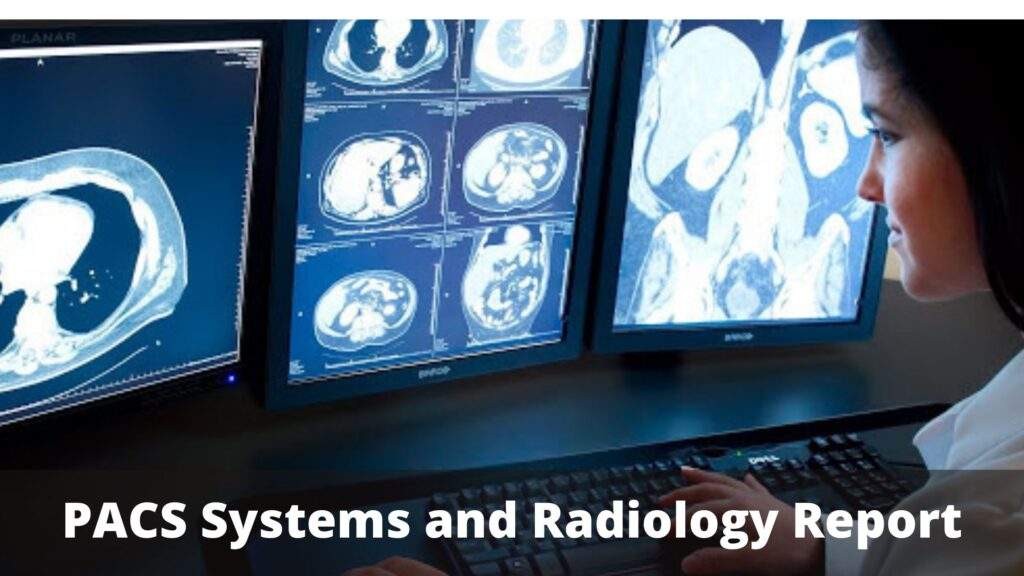
Brief Information About PACS Systems and Radiology Report

PACS Software (Picture Archiving and Communication System) is a medical imaging application that offers cost-effective recording, collection, management, dissemination, and medical pictures. Computerized images and reports can get electronically transmitted through PACS software. This removes the need to file, retrieve physically, and ship film jackets. It helps a healthcare institution record, archive, display, and exchange all sorts of images internally and externally.
DICOM (Digital Imaging and Communications in Medicine) is the standardized format for PACS image preservation and transition. DICOM helps PACS, Radiology Information Systems (RIS), and even more diagnostic imaging systems to link and relay data to systems in other medical facilities.
Most PACSs manage images from different diagnostic imaging instruments, including computed radiography (CR), magnetic resonance imaging (MR), nuclear medicine imaging, ophthalmology, positron emission tomography (PET), endoscopy (ES), mammograms (MG), computed tomography (CT), digital radiography (DR), histopathology, ultrasound (US), etc. Different kinds of image formats are always placed. Clinical areas beyond radiology, such as cardiology, oncology, orthopedics, and the laboratory, produce medical images that can get integrated into the PACS.
The PACS consists of four main components:
- The mode of imaging;
- A protected network for transmitting patient data;
- Workshops for analyzing and evaluating images;
- And archives for storing and retrieving images and reports.
PACS can provide timely and efficient access to images, explanations, and associated data in combination with currently offered and emerging web technologies. It shatters down the physical and time barriers associated with the extraction, allocation, and display of traditional film-based images.
PACS Systems: Benefits
- Hard Copy Substitution: PACS software substitutes hard copy-based tools for managing medical images, like movie records. With the declining price of digital storage, they have an increased cost and space benefit over film collections and immediate access to previous images at the same institution. Digital versions are referred to as soft copy.
- Distant Access: It widens conventional systems’ options by providing off-site watching and monitoring requirements (online learning, tele diagnosis). It allows clinicians in various geographical locations to concurrently access the same knowledge for teleradiology.
- Electronic Image Connectivity Network: It offers an electronic platform for X-ray image interfaces for other medical workflow applications such as Radiology Information System (RIS), Practice Management Software, Hospital Information System (HIS), Electronic Medical Record (EMR), and others.
- Radiology Process Management: Radiology employees use PACS to monitor patient tests’ workflow.
The specialty in radiology is an area of medicine with a rather particular interest in PACS applications. PACS radiology is commonly deployed with RIS. RIS is used to plan hospital visits and document the patient’s radiology records, where PACS focuses mainly on image storage and rehabilitation. It is not just the radiologists who have to be persuaded of PACS’s importance and cost-effectiveness, but also the referral of doctors and hospital managers.
Transforming Healthcare System
The healthcare system is transforming, and customized healthcare is becoming more relevant. From this point of view, each patient is unique. It is also of the highest significance to consider as much data as possible, including information derived from medical photographs. However, many radiologists are already stretched as they are. Introducing more details to the radiology reporting requires time and gives rise to additional questions from referral clinicians.
This will add to a significant increase in the workload during a working day that is still overcrowded. Thus, the critical question to be answered, particularly in this time of artificial intelligence (AI) in radiology, is how to consider and report as much data as possible without overburdening radiologists with much more tasks.
Interaction between radiologists and referral clinicians, like neurologists, is a critical aspect of the diagnostic work process. Communication can take place in different ways at different times-at the formal multidisciplinary team meeting (MDT) or even as a quick ask for advice in the corridor, and everything in between.
Improving this critical aspect of the diagnosis chain has a tremendous ability to reduce the massive workload of both the radiologists and the referral doctors while at the same time offering more information on the patient in the decision-making method. This is particularly the case where part of the report’s information collection and processing can be standardized.
Four Advantages of an Optimized Radiology Study
The radiology reporting can get streamlined in several ways, e.g., by introducing (more) structure to the text, integrating main images, providing (more) quantitative statistics, or displaying these data in clearly readable graphs. Many specialist radiological societies now advocate including various uniform elements to improve radiologist reporting and reduce inter-reader variance. However, studies on radiology should be more strengthened. This will come you realize the following benefits:
1.More effective contact with referral doctors using optimized radiology records
The format of every report is crucial to accurately communicating key observations while mitigating radiologists’ differences. Clinicians can quickly see the necessary data without needing to read the entire article. Also, placing the patient outcomes in perspective at one glance will speed up the data analysis. Still, it will also include comprehensive information that is useful for the diagnosis.
2.Improved coordination with MDT using optimized radiology reports
Multidisciplinary team sessions will be quite lengthy as multiple clinicians present as they all have a massive commitment to share. Imagine just requiring a few graphs, instantly providing the critical insights derived from medical scans, and accelerating information transfer during the MDT.
3.Refined communication of patients through optimized radiology reports
Radiology reporting can be complicated for a patient to understand. This is not unusual, as the file is not explicitly mentioned to patients. It aims to communicate the findings of radiology to the referral physicians. However, what if all the relevant parties can only use one file to interact with critical results? Radiologists, clinicians, and patients all would have access to the right data without anyone replicating it from one (digital) place to the next.
4.Improved basis for algorithm training utilizing radiology reports
The standardization of data collection has one significant added benefit: it can make an efficient algorithm much simpler. Ordered data is the secret to the practice of well-functioning AI algorithms. A specific and full recording of the data would allow the programmer to choose the data required for preparation. This is of the greatest priority as a lack of selection will induce the bias of the algorithm.


